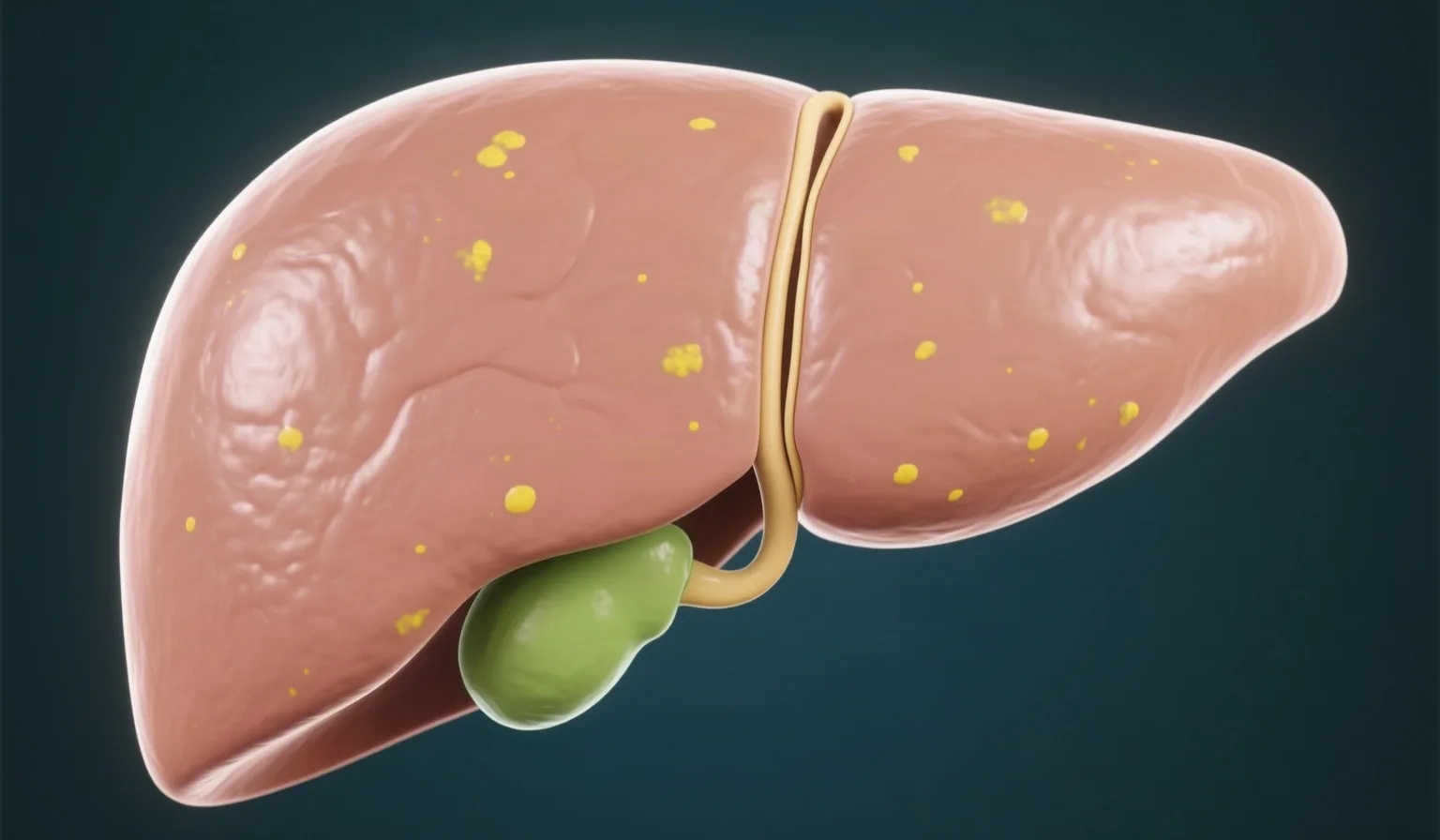
An upper abdominal ultrasound report often says “fatty infiltration of the liver, grade 1, 2 or 3.” The wording causes concern because it sounds like the staging of a disease. It is not. The grade describes how much fat the radiologist sees in the liver, not how much damage the organ has suffered.
This distinction is the most important piece of information for anyone holding such a report, because it determines whether further testing is needed and what kind.
What the ultrasound shows when the report mentions fatty liver
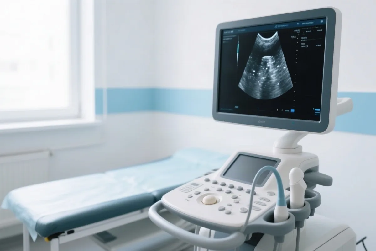
Fat changes the way ultrasound waves pass through the liver tissue. The more fat builds up in the liver cells (hepatocytes), the more echogenic – that is, the “whiter” – the liver appears, and the more the beam weakens as it travels toward the deeper parts.
The assessment is qualitative and depends on the examiner. Two radiologists may describe the same image differently, especially in the milder forms. That is why a grade should not be compared mechanically with the grade from a previous ultrasound done elsewhere.
The three grades on ultrasound
The classification is based on three features: the echogenicity of the liver compared with the cortex of the right kidney, how sharply the walls of the blood vessels inside the liver can be seen, and the visibility of the diaphragm.
Grade 1, mild steatosis
A mild, diffuse increase in the echogenicity of the liver. The walls of the portal vein and the diaphragm remain clearly visible. At this grade, ultrasound is least accurate, so mild steatosis may either be missed or overestimated.
Grade 2, moderate steatosis
A moderate increase in echogenicity, with reduced sharpness of the walls of the portal vein and of the diaphragm. This is the finding most often mentioned in reports.
Grade 3, severe steatosis
A marked increase in echogenicity, with little or no visualization of the walls of the portal vein, the diaphragm and the back (posterior) part of the right lobe. Here ultrasound is reliable, since its sensitivity increases as the percentage of fat rises.
Why the ultrasound grade is not a disease stage
This is where the most common misunderstanding arises. It is widely claimed that grade 1 means fat in 5 to 33% of liver cells, grade 2 in 34 to 66%, and grade 3 in more than 66%. These percentages come from the histological grading of steatosis, that is, from a biopsy, and they are not a definition of the ultrasound grades. Ultrasound does not measure percentages.
The practical takeaway: the grade shows how much fat there is, not whether there is inflammation or fibrosis. And the prognosis is determined by fibrosis, not by fat.
Steatosis, steatohepatitis and fibrosis
Three terms that are often confused, even though their severity is completely different.
- Steatosis: a simple buildup of fat in the liver cells. This is what ultrasound sees.
- Steatohepatitis: fat together with inflammation and damage to the cells. It cannot be diagnosed with ultrasound.
- Fibrosis: replacement of normal tissue with scar tissue. It is the factor that determines the prognosis, and it can lead to cirrhosis or hepatocellular carcinoma.
A liver with grade 3 steatosis and no fibrosis has a better prognosis than a liver with grade 1 steatosis and advanced fibrosis. The grade on its own does not answer the question “How serious is it?”
Which tests complement the ultrasound
Blood tests and the FIB-4 index
Liver enzymes alone are not enough to assess fibrosis. The 2024 EASL-EASD-EASO guidelines recommend the FIB-4 index as the first step. It is calculated from age, the transaminases AST and ALT, and the platelet count – values that are usually already available from a standard blood test.
A FIB-4 value below 1.3 makes advanced fibrosis unlikely. A value above 2.67 leads to a specialist assessment. Intermediate values, from 1.3 to 2.67, do not answer the question and call for a second test, usually elastography. In people over 65 years of age, the lower cutoff rises to 2.0, while in people under 35 the index is not considered reliable. These adjustments exist so that age, which is part of the formula, does not produce false-positive or false-negative results.
Elastography and CAP
Elastography measures the stiffness of the liver, which gives an approximate picture of fibrosis. On the same device, the CAP (controlled attenuation parameter) estimates the amount of fat quantitatively. In the table of the 2024 EASL guidelines, the cutoff values for CAP are 248 dB/m for S1, 268 dB/m for S2 and 280 dB/m for S3. This is an objective measurement, unlike the grading of a standard ultrasound.
Magnetic resonance imaging (MRI)
The MRI-PDFF technique measures the percentage of liver fat accurately. Because of its cost and availability, it is used mainly in clinical studies and in selected cases.
Liver biopsy
It remains the reference method for distinguishing simple steatosis from steatohepatitis and for staging fibrosis, but it is not needed in most cases. It is reserved for unclear or advanced cases.
When the grade changes management
In practice, the grade serves as a prompt for further evaluation, not as a guide to treatment. The tests that follow are the same whether the report says grade 1 or grade 3: assessment of cardiometabolic risk factors, blood tests, calculation of FIB-4 and, where indicated, elastography.
What changes is how urgently lifestyle changes are needed and how intensive the follow-up should be, especially when type 2 diabetes, obesity, high blood pressure or abnormal blood fats (dyslipidemia) are also present.
Is fatty liver reversible?
Simple steatosis is largely reversible. According to the 2024 EASL-EASD-EASO guidelines, in adults with overweight or obesity, weight loss should aim for a sustained reduction of 5% or more to reduce liver fat, 7 to 10% to improve inflammation, and 10% or more to improve fibrosis. In people of normal weight, the target is set at 3 to 5%. Once fibrosis has developed, it regresses with more difficulty and more slowly.
The key measures are gradual weight loss, a Mediterranean-style diet that limits sugar and processed foods, regular aerobic exercise, and cutting down on or avoiding alcohol. You can read more about the overall picture of the condition on the page about fatty liver disease.

From NAFLD to MASLD
In June 2023, the international nomenclature changed. The term non-alcoholic fatty liver disease (NAFLD) was replaced by metabolic dysfunction-associated steatotic liver disease (MASLD), and non-alcoholic steatohepatitis (NASH) was renamed MASH. The change does not affect the patient’s diagnosis but the way the disease is classified, with an emphasis on metabolic factors rather than on the absence of alcohol.
Frequently asked questions
Sources
- EASL – EASL-EASD-EASO Clinical Practice Guidelines on the management of metabolic dysfunction-associated steatotic liver disease (MASLD). https://www.journal-of-hepatology.eu/article/S0168-8278(24)00329-5/fulltext
- National Library of Medicine – Relationship Between Greyscale Ultrasound Grading of Hepatic Steatosis and Attenuation Imaging. https://pmc.ncbi.nlm.nih.gov/articles/PMC9038208/
- National Library of Medicine – Pathological Evolution and Internal Medicine Management of NAFLD in the Era of MASLD. https://www.ncbi.nlm.nih.gov/pmc/articles/PMC12306519/
- Australasian Journal of Ultrasound in Medicine – Hepatic steatosis: Qualitative and quantitative sonographic assessment in comparison to histology. https://onlinelibrary.wiley.com/doi/full/10.1002/ajum.12381
Book your appointment
If your ultrasound showed fatty liver, an evaluation by a gastroenterologist helps to assess the condition of the liver and to decide on the appropriate tests. To schedule your visit, please contact Dr. Thomaidis.
Send your request
Fill in your details and we will contact you to schedule your appointment.

About the doctor
Dr. Thomas Thomaidis, MD, PhD – Interventional Gastroenterologist. Associate Professor of Gastroenterology specializing in diagnostic and therapeutic endoscopy, member of international scientific societies (ASGE, DGVS, Hellenic Society of Gastroenterology). See the full CV.
This article is for information purposes only and does not replace personalized medical advice, diagnosis or treatment. For your own symptoms and their treatment, please consult a doctor.
Content last updated: October 2026 (12/10/2026).