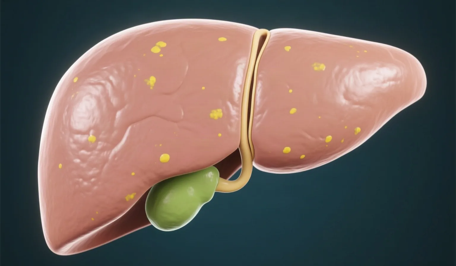
A cyst on the pancreas is found by chance more and more often, on a CT or MRI scan done for another reason. The finding often causes concern, but it is quite common. In adults without symptoms it is found in 2.4% to 24.3% of cases, a figure that rises with age: from 0.5% under the age of 40, to 11% between the ages of 70 and 79, and to 37% after 80.
Pancreatic cysts are not all the same. Some are benign, while others may change over the years and are therefore monitored. The first and most important step is to clarify what type of cyst it is, because this determines whether anything more will be needed.
Types of pancreatic cysts
| Type of cyst | What it is | What it usually means |
|---|---|---|
| Pseudocyst | A collection of fluid related to pancreatitis | It is not a neoplasm; when it causes no symptoms, it does not need surveillance |
| Serous cystadenoma (SCN) | A neoplasm that is more common in women, usually in the body or tail of the pancreas | Considered benign; surveillance for 1 year and after that only if symptoms appear |
| Intraductal papillary mucinous neoplasm (IPMN) | A mucinous cyst of the pancreatic duct system, involving the main duct, the side branches or both | The main-duct type has a high risk of malignancy and is removed when the patient is fit for surgery; the side-branch type is usually monitored |
| Mucinous cystadenoma (MCN) | Mainly in middle-aged women, usually in the tail of the pancreas, with no communication with the pancreatic duct | Surgical removal or surveillance is decided individually, depending on the patient and the size of the cyst. |
| Solid pseudopapillary neoplasm (SPN) | A rare neoplasm | Surgical removal is recommended. |
| Cystic neuroendocrine tumor | Rare; cytology with endoscopic ultrasound supports the suspicion, while the definitive diagnosis is histological | Surgery when it is larger than 20 millimeters; surveillance for smaller ones without symptoms |
Telling the types apart is not always easy. The accuracy of MRI in identifying the type of cyst ranges from 40% to 95%, which is why an additional test is needed in some cases. An example from the practice is the case in which cysts that were first thought to be pseudocysts turned out to be IPMNs on endoscopic ultrasound.
Symptoms are usually absent
Many cysts are found in people without symptoms.
However, there are findings that need prompt assessment:
- jaundice, meaning yellowing of the skin and eyes, caused by the cyst,
- an episode of acute pancreatitis,
- new-onset diabetes in a patient with a known cyst,
- a raised CA 19-9 marker in the blood.
A cystic lesion can itself be the cause of an episode of acute pancreatitis.
The tests that are needed
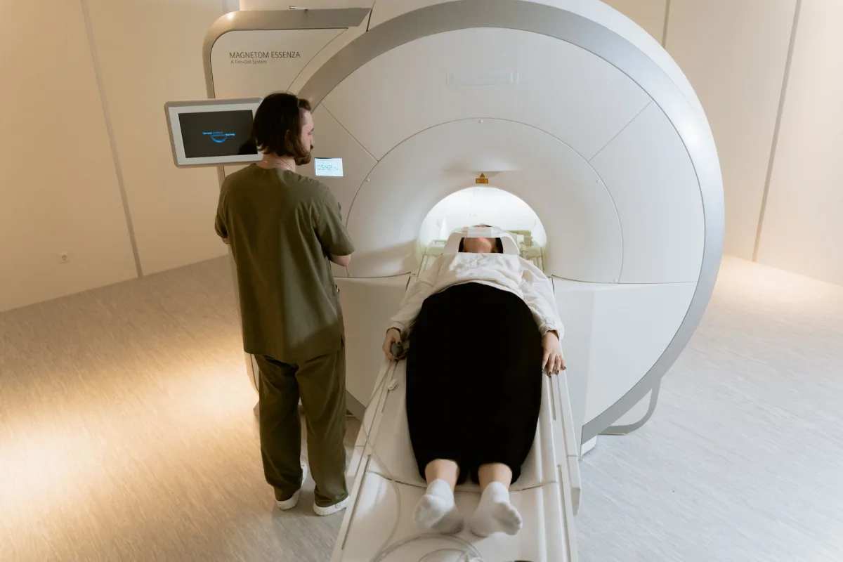
Magnetic resonance imaging with cholangiopancreatography (MRCP). It is the test of choice both for characterizing and for monitoring a cyst. It does not use ionizing radiation, which matters when surveillance lasts for years. It also shows better whether the cyst communicates with the pancreatic duct and whether it has internal septations, meaning thin walls that divide it into smaller spaces, or a nodule, meaning a small solid growth in its wall.
CT scan. It is useful when it matters to detect calcifications, meaning calcium deposits, for example to distinguish a pseudocyst in a patient with chronic pancreatitis. It is also used when malignancy is suspected and the involvement of blood vessels or the spread of the disease needs to be assessed.
Endoscopic ultrasound. Endoscopic ultrasound complements the other tests and is recommended when the cyst has clinical or imaging features of concern, either at the initial work-up or during surveillance. It shows the wall, the septations, the nodules and the communication of the cyst with the main pancreatic duct. With an intravenous contrast agent, contrast-enhanced endoscopic ultrasound assesses wall nodules better.
Fine-needle aspiration (EUS-FNA). During endoscopic ultrasound, fluid can be taken from the cyst to measure carcinoembryonic antigen (CEA) and pancreatic enzymes (amylase, lipase) and for cytology. The combination of CEA, lipase and cytology gives the best distinction between mucinous and non-mucinous cysts. A CEA level equal to or greater than 192 ng/mL is found more often in mucinous cysts, while amylase below 250 U/L makes a pseudocyst very unlikely. Cytology is very reliable when it is positive, but the sample is often not enough for a safe conclusion.
Aspiration is done only when the result is expected to change management, and not when the diagnosis is already clear or there is already an indication for surgery. Complications are rare, about 3.4%, and the vast majority are mild. Dr. Thomaidis performs endoscopic ultrasound and tissue sampling with EUS-FNA and EUS-FNB.
Features that change the outlook
For IPMN-type cysts, features have been defined that increase the likelihood of high-grade dysplasia, meaning precancerous changes in the cells, or malignancy.
Strong indications for surgical assessment:
- jaundice caused by the lesion,
- a wall nodule that enhances with contrast and measures 5 millimeters or more,
- a solid mass,
- cytology positive for high-grade dysplasia or malignancy,
- dilation of the main pancreatic duct to 10 millimeters or more.
Relative indications, which lead to closer surveillance or a discussion about surgery:
- growth of 2.5 millimeters or more per year,
- CA 19-9 above 37 U/mL, without jaundice,
- dilation of the main duct between 5 and 10 millimeters,
- a cyst size of 30 millimeters or larger,
- new-onset diabetes,
- acute pancreatitis caused by the cyst,
- a nodule that enhances with contrast and is smaller than 5 millimeters.
The decision is individualized for each patient. When the patient can no longer undergo surgery because of their general condition, surveillance is not continued.
Surveillance
Most cysts that need surveillance are monitored with MRI, while endoscopic ultrasound is used in selected cases.
- IPMN without indications for surgery: a follow-up scan at 6 months in the first year and then every year.
- IPMN with relative indications, or in older patients or patients with serious other illnesses: a follow-up scan every 6 months.
- Small cysts of uncertain type, under 15 millimeters, without worrying features: a follow-up scan after 1 year and, if they stay stable for 3 years, every 2 years.
New-onset diabetes or growth of 5 millimeters or more per year leads to a follow-up scan every 6 months. Surveillance continues as long as the patient could undergo surgery if it were needed. For side-branch IPMNs under 20 millimeters that remain unchanged after 5 years of surveillance, the newer international guidelines also accept stopping it. The decision takes into account the patient’s general condition and life expectancy. The schedule is set by the doctor for each patient individually.

When surgery is needed
Only some cysts eventually need surgery. Surgery is discussed when the strong indications mentioned above are present. It is also discussed for main-duct IPMN in a patient who is fit for surgery, for a mucinous cystadenoma or a side-branch IPMN with worrying features, for a solid pseudopapillary neoplasm and for a cystic neuroendocrine tumor larger than 20 millimeters. A serous cystadenoma is removed only when it presses on nearby organs, such as the bile duct or the stomach, and causes symptoms.
Because pancreatic operations carry significant morbidity, they are performed in specialized pancreas centers, with a multidisciplinary approach, after weighing the benefit against the risk for the individual patient.
Frequently asked questions
Book your appointment
Send your request
Fill in your details and we will contact you to schedule your appointment.
Sources
- The European Study Group on Cystic Tumours of the Pancreas, European evidence-based guidelines on pancreatic cystic neoplasms. Gut 2018;67(5):789–804. https://pmc.ncbi.nlm.nih.gov/articles/PMC5890653/
- Canakis A, Lee LS, State-of-the-Art Update of Pancreatic Cysts. Digestive Diseases and Sciences 2022;67(5):1573–1587. https://pmc.ncbi.nlm.nih.gov/articles/PMC9142439/
- Ma T, Liang T, Surveillance of non-resected branch-duct intraductal papillary mucinous neoplasms: is a simplified algorithm justified? HepatoBiliary Surgery and Nutrition 2025;14(1):136–139. https://hbsn.amegroups.org/article/view/133274/html

About the doctor
Dr. Thomas Thomaidis, MD, PhD – Interventional Gastroenterologist. Associate Professor of Gastroenterology specializing in diagnostic and therapeutic endoscopy, member of international scientific societies (ASGE, DGVS, Hellenic Society of Gastroenterology). View the full CV.
This article is for informational purposes only and does not replace individualized medical advice, diagnosis or treatment. For your own case and your surveillance plan, please consult a doctor.
Content last updated: November 2026 (18/11/2026).