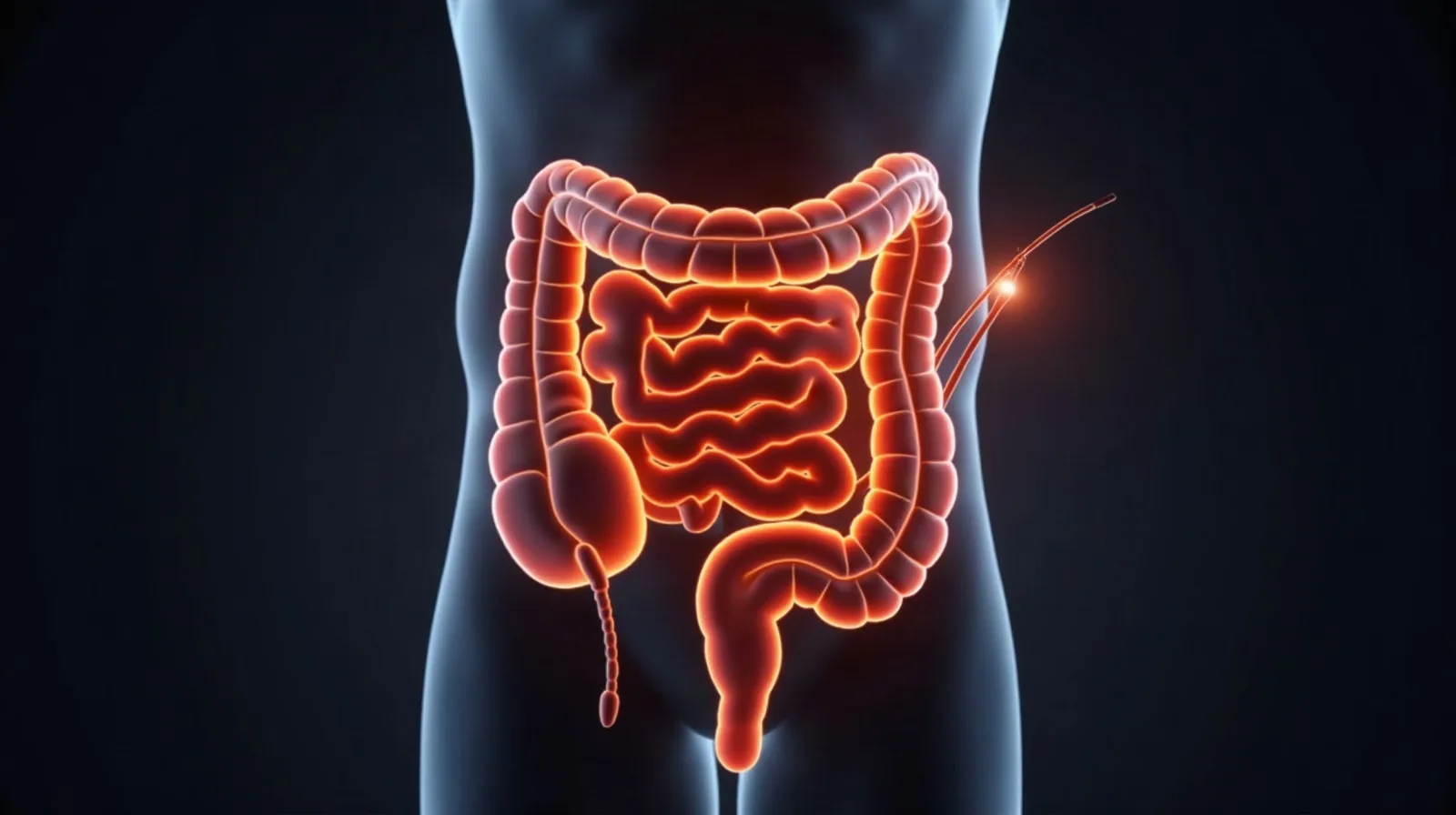
ESD of a large polyp in the rectum involving the dentate line
Cases from practice
The technique is applied throughout the digestive tract. Examples include a rectal resection, a 4 cm lesion of the prepyloric antrum and an oesophageal resection. Each location brings its own difficulty, from gastric motility to the thinness of the duodenal wall.
Why the dentate line changes the difficulty
The dentate line marks the transition from rectal mucosa to anal canal, and below it the tissue is richly supplied with somatic nerves. Dissection that crosses this boundary is felt by the patient in a way that rectal dissection is not, and it demands both different anaesthetic planning and a more deliberate technique.
What is being protected
Beyond the lesion itself, the aim is continence. Surgery for an extensive lesion at this level can mean resection of bowel and, in low positions, a permanent stoma. Removing the lesion from within leaves the sphincter mechanism untouched, which for the patient is the difference between a procedure and a change of life.
Lesions at the dentate line
The rectum is one of the sites where endoscopic removal offers the greatest benefit. For an extensive lesion the alternative is surgical resection of a bowel segment, and with low lesions a stoma may be required. ESD preserves the organ intact. Involvement of the dentate line adds difficulty, since the area is richly innervated and demands careful technique.
After the resection
The specimen is sent for histology, and it is that report rather than the endoscopic appearance that determines the next step. Surveillance intervals are set by guideline and individualised per case; they are set out in detail on the page for colonoscopy.
What the specimen must show
For a lesion at this level the pathology report answers two questions that determine everything that follows: whether the deep margin is clear, and whether invasion is confined to the mucosa. A lesion crossing into the submucosa carries a risk of lymph node spread that endoscopic removal cannot address, and that finding, not the appearance at the time, is what sends a patient to surgery.
Frequently asked questions
Sources
- European Society of Gastrointestinal Endoscopy, Post-polypectomy colonoscopy surveillance: ESGE Guideline Update 2020. https://doi.org/10.1055/a-1185-3109
Content last updated: September 2026 (22/09/2026).